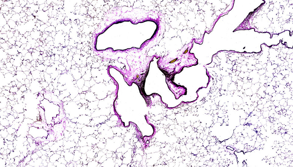
Verhoeff Van Gieson Staining: A Valuable Tool in Microscopy
For trend-aware readers interested in microscopy and histology, Verhoeff Van Gieson (VVG) staining is a technique that has gained significant attention. This staining method is primarily used to differentiate between collagen and elastin, two crucial proteins found in connective tissue. The VVG stain is especially useful in diagnosing and studying various diseases that affect the cardiovascular system, lungs, and skin. With its ability to highlight elastin, a key component of elastic fibers, the Verhoeff Van Gieson stain provides valuable insights into tissue structure and function.
What is Verhoeff Van Gieson Staining?
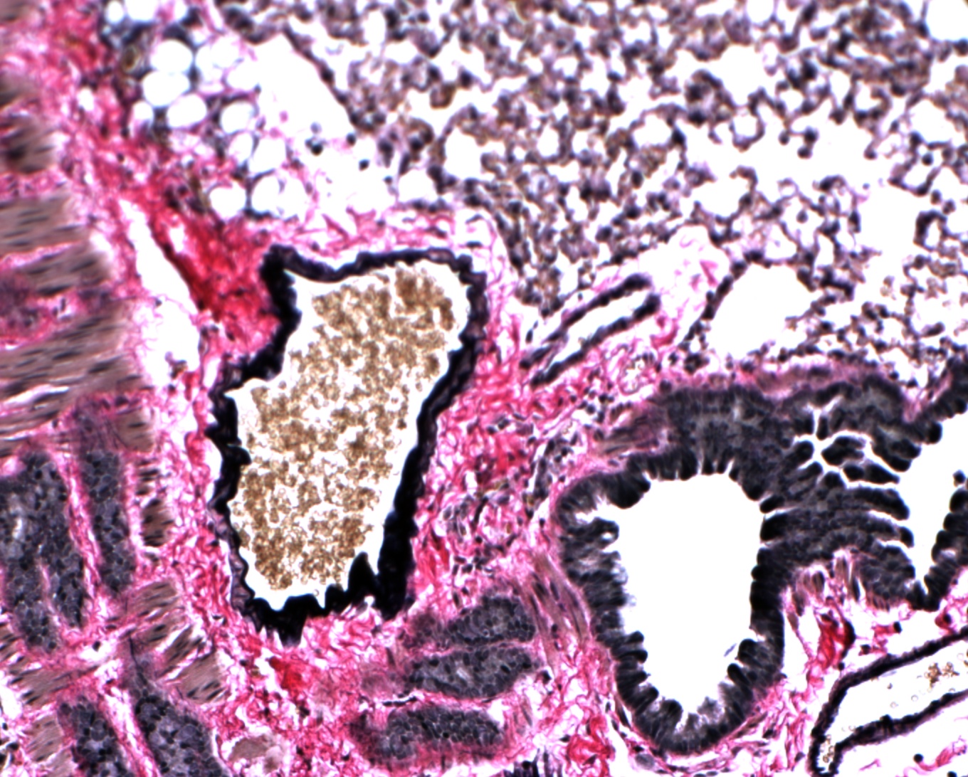
Verhoeff Van Gieson staining is a histological technique used to visualize elastin, a protein that gives tissues their elastic properties. The stain consists of a combination of dyes, including Verhoeff's iodine and Van Gieson's picro-fuchsin, which selectively bind to elastin, allowing it to be distinguished from other tissue components. This technique is particularly useful in the diagnosis of diseases such as atherosclerosis, where elastin degradation plays a significant role. The VVG stain has undergone various modifications over the years, but its core principle remains the same.
How Does Verhoeff Van Gieson Staining Work?
The Verhoeff Van Gieson staining process involves several steps, including tissue fixation, sectioning, and staining. The tissue sample is first fixed in a solution to preserve its structure, then sectioned into thin slices. The sections are then subjected to the VVG stain, which binds to the elastin present in the tissue. The resulting stained tissue can be viewed under a microscope, allowing researchers and clinicians to visualize the distribution of elastin. 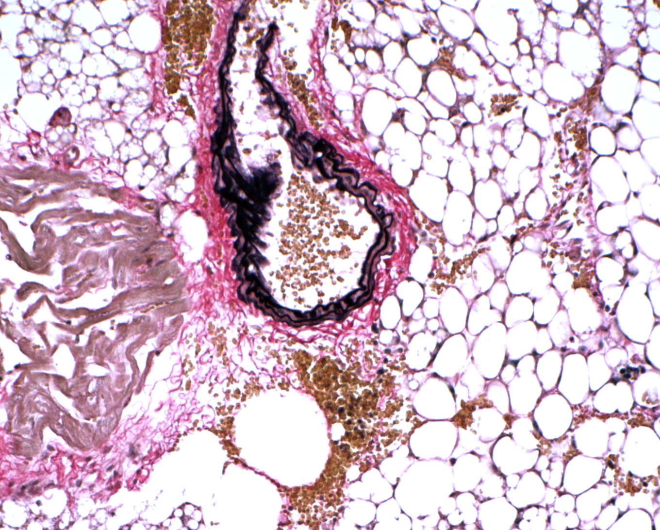
What Are the Advantages and Limitations of Verhoeff Van Gieson Staining?
The Verhoeff Van Gieson stain has several advantages, including its ability to selectively stain elastin, making it an invaluable tool in the diagnosis of diseases that affect elastic fibers. Some of the key benefits of VVG staining include:
- High sensitivity and specificity for elastin detection
- Ability to distinguish between collagen and elastin
- Relatively simple and cost-effective technique
- Potential for background staining, which can obscure elastin visualization
- Requires proper tissue fixation and sectioning to produce optimal results
- May not be suitable for all types of tissue samples
What Are the Implications of Verhoeff Van Gieson Staining in Research and Diagnosis?
The Verhoeff Van Gieson stain has significant implications in research and diagnosis, particularly in the field of cardiovascular disease. By visualizing elastin degradation, researchers can gain insights into the mechanisms underlying atherosclerosis and other diseases. The VVG stain can also be used to monitor the effectiveness of treatments aimed at preventing or reversing elastin degradation. 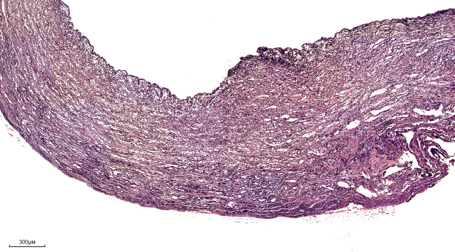
Conclusion
In conclusion, the Verhoeff Van Gieson stain is a valuable tool in microscopy and histology, providing a means to visualize elastin and study its role in various diseases. While it has its limitations, the VVG stain remains a widely used technique in research and diagnosis. As our understanding of elastin and its functions continues to grow, the importance of Verhoeff Van Gieson staining will only continue to increase, making it an essential technique in the field of microscopy and beyond.
VitroView™ Verhoeff Van Gieson Elastin Stain Kit
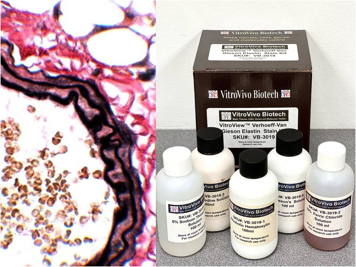
VitroView™ Verhoeff Van Gieson Elastin Stain Kit
VitroView™ Verhoeff Van Gieson Elastin Stain Kit
VitroView™ Verhoeff Van Gieson Elastin Stain Kit
Verhoeff Van Gieson Stain | Elastic & Collagen Fiber Detection | IHisto
Verhoeff Van Gieson Stain | Elastic & Collagen Fiber Detection | iHisto